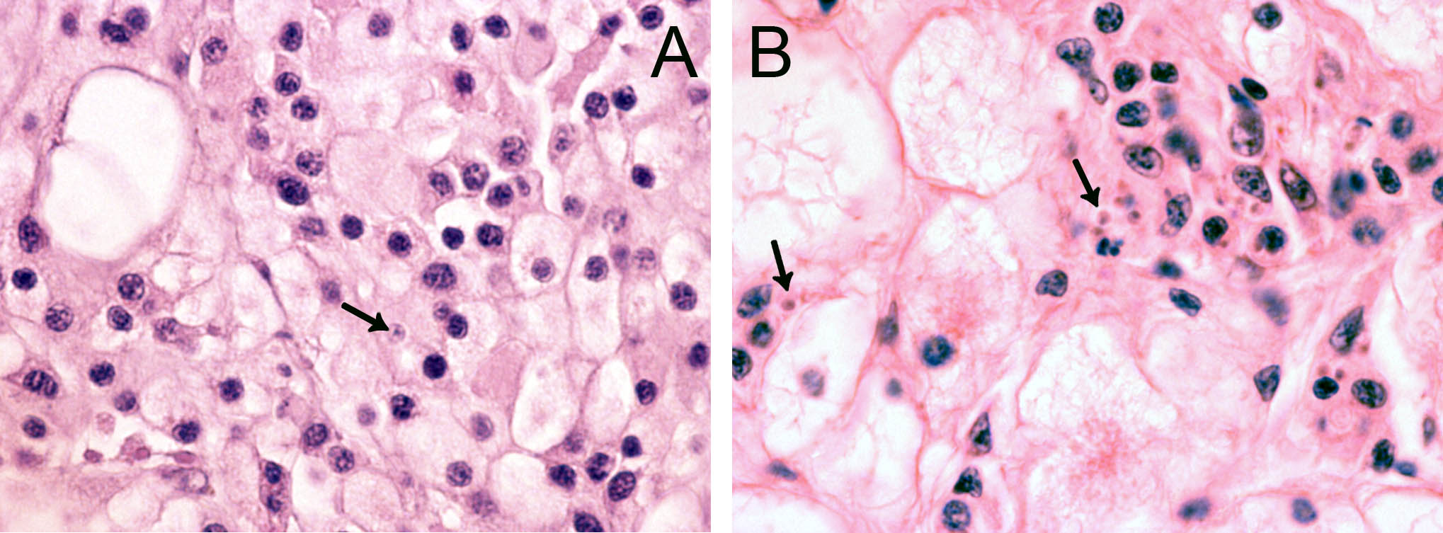
Figure
1. A: Section
showing connective/storage tissues from a flat oyster, Ostrea
edulis
from Langestrand near Arendal, june 2009. Numerous haemocytes are
present in the tissue. An un-identified cell is marked with an arrow.
B:
Same tissue area in a reference section from a flat oyster infected
with Bonamia
ostreae ,
provided by EURL. Several B.
ostreae are
visible (arrows), both freely and inside haemocytes. Both images have
the same magnification.