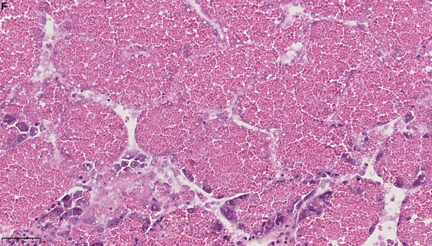
Figure 1. E) Normal tubules filled with lysosomes in the digestive cells; basophilic cells (yellow arrow); digestive cell (black arrow); the lysosomes appear to be released into the lumen in some tubules (Asterix), F) Autolysis of the entire structure of DD. Note the disappearance of the basal membrane and the basophilic cells. HES stained. NDP view 2, 40X.