The surveillance and control programme for bonamiosis and marteiliosis in European flat oysters, Ostrea edulis, and blue mussels, Mytilus sp. in Norway in 2018
Overvåkings- og kontrollprogram for bonamiose og marteiliose i flatøsters, Ostrea edulis, og blåskjell, Mytilus sp. i Norge i 2018Rapport NOK østers 2018
Overvåkingsprogrammet for sykdommene bonamiose og marteiliose i flatøsters og blåskjell utføres av Havforskningsinstituttet på oppdrag fra Mattilsynet. Det ble hentet skjell fra fire ville bestander, to blåskjellanlegg og ett østersanlegg. Høsten 2018 ble det for første gang inkludert prøver fra blåskjell fra Trøndelag. Prøver ble samlet inn i April/Mai og i Oktober, når prevalensen av parasittene Bonamia spp. og Marteilia spp. er høyest i smittede bestander. Det ble ikke observert unormal dødelighet verken vår eller høst. Bonamia ostreae / B. exitiosa ble ikke påvist. Resultatene kan danne bakgrunn for en søknad om etablering av fristatus for Bonamia spp. i norsk flatøsters.
Det er kommet inn en rekke rapporter om at blåskjell «forsvinner» mange steder langs kysten. Årsakene til dette er ikke kjent. Parasitten Marteilia sp. ble imidlertid for første gang påvist I blåskjell, Mytilus edulis, på Bømlo i 2016. Denne påvisningen er fulgt opp med en utvidet prøvetaking i HI-prosjekt Blåskjelldødelighet (83737-04) i 2017 og 2018. Det er gjort smittestudier som gir informasjon om smittetidspunkt og -forløp. Prøver samlet inn i Juli 2018 tyderpå at parasitten også kan finnes i den nedlagte østerspollen i Espevik, Tysnes.
Resultatene viser at infeksjonen med Marteilia sp. er begrenset til blåskjell. Østers fra den samme lokaliteten blir ikke smittet. Genetiske studier av Marteilia spp. fra England, Sverige og Norge (Bømlo) er inkludert i en studie som er gjort i EU-prosjektet VIVALDI. Marteilia sp. fra disse områdene er ulik Marteilia refringens som forårsaker sykdom hos flatøsters og er foreslått gitt navnet Marteilia pararefringens. Det ser således ut til at Marteilia refringens og Marteilia pararefringens sp. nov. er ulike arter med ulike vertsarter (hhv østers og blåskjell). På bakgrunn av funnet av M. pararefringens er det viktig å skaffe mer informasjon om helsestatus hos blåskjell langs hele kysten. Dette kan gjøres gjennom en utvidet helseovervåking, eventuelt gjennom en ny modell hvor overvåking og helsekontroll kobles, for å generere mer data.
Summary
The surveillance programme is carried out by the Institute of Marine Research according to a contract with the Norwegian Food Safety Authority. Samples were collected from four wild beds, two mussel farms and one oyster farm. Samples were collected in April/May and in October, in order to be able to detect Bonamia sp. and Marteilia sp. during the periods when the potential prevalence could be at the highest. No abnormal mortalities were observed in oyster populations during the surveillance. Bonamia ostreae / B. exitiosa were not detected. The results may be used as a background for an application for disease free status for Norwegian flat oysters.
There have been several reports on mortality or “disappearance” of mussels along the Norwegian coast. The reason(s) for the mortalities have not been determined. However, the parasite Marteilia sp. was detected for the first time in mussels, Mytilus edulis at Bømlo, western Norway, collected during the surveillance programme in 2016. This has been followed up with an extended survey in the IMR research project Mussel mortalities (83737-04). We have performed transmission experiments that identify the time period of infection and the progress of the infection in mussels. Analyses of samples collected in July 2018 indicate that the parasite may be present in an abandoned oyster lagoon at Espevik, Tysnes.
The results from the research project indicate that the Marteilia sp detected is limited to mussels. Flat oysters at the same site do not become infected. This is relevant to the listing of susceptible hosts for Marteilia spp. A genetic study of Marteilia spp. from the UK, Sweden and the present site at Aga has been included in a study in the EU-project VIVALDI. The name Marteilia pararefringens has been proposed, and there is strong evidence that Marteilia refringens and Marteilia pararefringens sp. nov. are distinct parasites of bivalves and have different European distributions. After the detection of M. pararefringens in mussels, it is important to obtain more data from mussels along the Norwegian coast. Mussels from Trøndelag in 2018 represented the first samples from mussels north of Bergen. In order to obtain a better set of data, we propose an extended surveillance that could be obtained through a revised surveillance progamme combined with a new model for health control in mollusk farms.
Updated 12/04/2023: Missing text in several of the sections has been added.
1 - Introduction
Norwegian populations of European flat oysters, Ostrea edulis, have been considered free from notifiable diseases. In 2006, microcells resembling the oyster parasite Bonamia sp. were observed during histopathological examination of tissue specimens of flat oysters, Ostrea edulis from the Arendal area, southern Norway. In 2008, the EU reference laboratory received samples from the Norwegian Veterinary Institute, and reported one Bonamia sp. in a haemocyte from one oyster. By real-time PCR, positive results were obtained from two oysters in one triplicate sample. The parasite has however never been detected during examination carried out by the Norwegian Veterinary Institute or Institute of Marine Research. Since 2009, more than 2 700 oysters have been examined by histology and/or PCR, all with negative results. The situation has thus been stable since 2006 (see 2017 report and Mortensen et al. 2016).
The surveillance programme for bonamiosis and marteiliosis in European flat oysters, Ostrea edulis, and blue mussels, Mytilus sp. is carried out by the Institute of Marine Research according to a contract with the Norwegian Food Safety Authority. The programme was revised in 2015. The parasite Marteilia sp. was detected in mussels at one site in 2016, and we increased the effort in 2017 and 2018 in order to study this case – including distribution and parasite life cycle.
This report gives a brief overview of the present situation, results from 2018 and suggestions for the 2019 sampling.
2 - Material and methods
The surveillance was performed according to EU directive 2006/88 and Decision 2015/1554. The sampling strategy, including wild beds and bivalve farms in operation, was revised in January 2015, and used as a background for the targeted surveillance also in 2018.
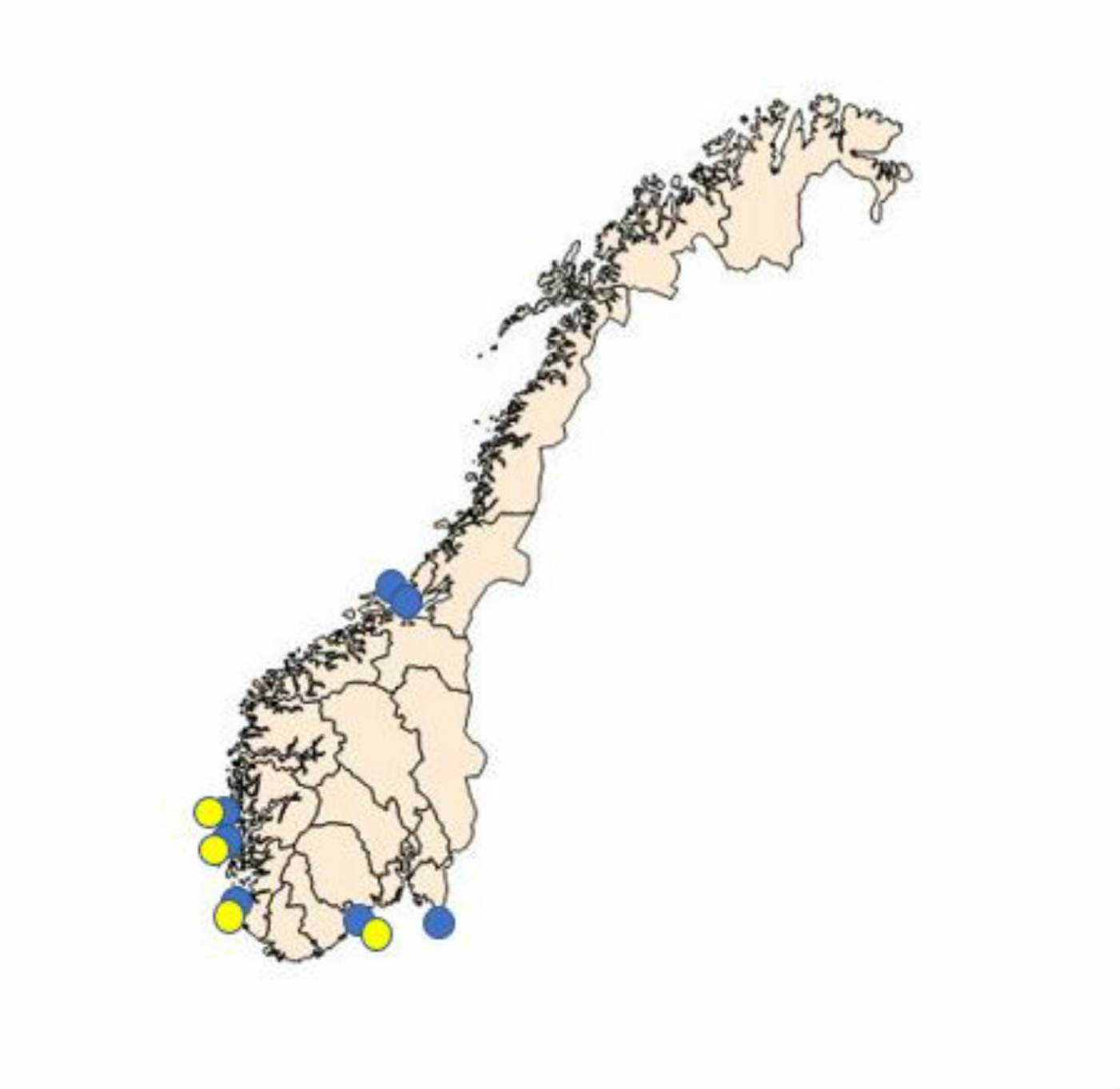
Sampling periods were defined according to the periods when the highest prevalence of Bonamia ostreae and Marteilia sp. (sporulating stage) have been detected in the northernmost areas where they have been detected (Engelsma et al. 2010; A. Alfjorden pers.comm). The selected sampling sites are shown in Figure 1 and listed in Table 1.
At Hafrsfjord and Langestrand, oysters and mussels were collected by skin-diving or wading in April and October and transported to the Institute of Marine Research (IMR) in Bergen. At Sveio, oysters and mussels were collected by the shellfish farmer and sent to IMR Bergen by over-night mail (Table 1). From Ytre Hvaler,
Østfold, mussels were collected and sent to IMR by over-night mail. At Aga, mussels and oysters were collected in the poll and brought directly to the laboratory in Bergen. Additional samples were taken from fauna. Samples from plankton and mussels were also collected in two other oyster lagoons; Innerøypollen (Halhjem) and Espevikpollen (Tysnes). In 2018, mussels from the two dispatch centers processing mussels in Trøndelag were included for the first time. Trøndelag is the main production area for mussels in Norway.
All oysters and mussels were processed at the IMR laboratory in Bergen, according to standard methodology, and under ISO 17025 QA. Briefly; Histology was performed using dorso-ventral cross sections, fixed in Davidson’s fixative, embedded in paraffin, sectioned at 3µm, stained with Hematoxylin Eosin Saffron (HES), mounted with a cover slip and observed at 100 to 1000 x magnification. Samples for PCR were fixed in ethanol. DNA was extracted from ethanol fixed digestive gland tissue from mussels from Aga. Marteilia refringens detection and typing was done with by Polymerase Chain Reaction, and as described by Le Roux et al (2001). Samples where (Bonamia-like) microcells were observed by microscopy were forwarded to real-time PCR as described by Corbeil et al 2006.
Healthy mussels were deployed at the infected site in May and October. Thirty mussels were sampled every six weeks until April 2019 and analyzed by histology and PCR.
Fauna samples and additional samples from mussels and oysters were collected in May, July and October. Samples were processed and analyzed by PCR as described above, at IMR or CEFAS, UK.
Figure 1. Sampling of flat oysters, Ostrea edulis, and blue mussels, Mytilus edulis in the surveillance programme for Bonamia sp. and Marteilia sp. Yellow circles indicate the sampling sites for flat oysters. Blue circles indicate the sampling sites for mussels.
Sampling site
Oysters
Mussels
Spring
Autumn
Ytre Hvaler, Østfold
30
Langestrand, Agder
150 (20) 30
30
NB: delayed reading of slides. Histo & PCR (30) spring
Hafrsfjord, Rogaland
30
30
30
Histo & PCR (30) spring
Sveio, Hordaland
30
Aga Bømlo, Hordaland Rogøysund, Bømlo
30
30
10
Additional study of Marteilia pararefringens in mussels at Aga, see text
Innerøyen, Hordaland
30
July, PCR
Espevik, Tysnes, Hordal.
30
July, PCR
Åfjord, Trøndelag
30
Rissa, Trøndelag
30
Table 1. Sampling and surveillance of flat oysters (Ostrea edulis) and mussels (Mytilus sp.) in 2018.
3 - Results
Bonamia spp. was not observed in any sample during 2018.
Langestrand, Aust-Agder.
The site was inspected by skin diving in May 2018. Dense oyster beds were observed down to approximately four-meter depth, with several cohorts present. There was no sign of abnormal mortality. Few adult Pacific oysters (Crassostrea gigas) were observed between the flat oysters. During sampling, Pacific oyster spat were observed on and in-between flat oyster shells and on pebbles in the inter-tidal zone.
Oysters: The reading of slides from oysters is delayed (see Table 1). During examination, gross morphology of shells and soft parts appeared normal. In the first 20 slides Bonamia ostreae / B. exitiosa or microcells resembling Bonamia spp. were not detected.
Mussels appeared normal, however most specimens had green pustules, presumably representing infections with the parasitic algae Coccomyxa parasitica (see Mortensen et al. 2005). Marteilia sp. was not observed.
Hafrsfjord, Rogaland
Samples were collected in May and October (Table 1). Sampling was performed by Knut Magnus Persson in conjunction collection of flat oysters for a re-stocking programme off the coast of The Netherlands. No sign of abnormal mortality was reported. A few adult Pacific oysters (Crassostrea gigas) were observed between the flat oysters on shallow water. Bonamia sp. or Marteilia sp. were not observed in mussels or oysters. Bonamia PCR of oysters were negative. During examination of the flat oysters, perforations due to Polydora sp. infestations were observed in shells from most of the oysters. Gross morphology of soft parts appeared normal. Rickettsia-like organisms (RLO’s) were observed in three oysters. Haemic neoplasia was observed in one oyster. The condition of the mussels was variable. Focal inflammations that may represent infections with C. parasitica was observed in five mussels.
Sveio, Hordaland
Oysters sampled in spring appeared healthy and in good condition. The dispatch centre at Sveio (Sunnhordland havbruk) has been closed, and the site was excluded from the surveillance programme after the spring sampling.
Ytre Hvaler, Østfold
Mussels from Ytre Hvaler (Papperhavn) were in variable condition. Microcells were observed in connective tissues, in some cases associated with inflammations. Green spots were observed in mantle tissues of all mussels, and the infestations were considered infections with C. parasitica. Most mussels were moderately infected with trematode cysts. Un-known microcells were observed inside cells in the stomach epithelium. Histological slides and tissue fixed in ethanol was forwarded to the Community Reference Laboratory for further investigation.
Aga, Bømlo, Hordaland: Studies on the Marteilia infection
Oysters: The condition index of the oysters was variable. Haemic neoplasia was observed in one oyster, and Rickettsia-like colonies (RLO’s) were observed in the digestive epithelia of three oyster. Marteilia sp. was not observed and the Marteilia-PCR (spring and autumn) were negative.
Mussels: Marteilia pararefringens sp. nov was detected in mussels in the Aga poll in all samplings. A focused study on the infection cycle in mussels was carried out with sampling every 6 weeks from May 2018 to April 2019. The results from this study will be presented separately, as a master thesis, later as a publication. Mussels from Rogøysund (receiving oyster spat from Aga and also used in this study as negative control), were negative.
Analyses of mussels from two other lagoons; Innerøyen and Espevik
PCR performed on mussels from Innerøypollen was negative. However, when PCR was conducted on mussels from Espevikpollen (Tysnes), two mussels were Marteilia-positive.
Åfjord, Trøndelag
Mussels from Åfjord appeared in good condition. No abnormal finding was recorded.
Rissa, Trøndelag
Mussels from Rissastrømmen appeared in good but variable condition. Green colonies (C.parasitica) was observed in the mantle of two mussels. No other abnormal finding was recorded.
4 - Discussion and conclusions
Examination of flat oysters
The wild flat oyster populations examined appears healthy, with a normal reproductive cycle pattern. Haemic neoplasia and the presence of intracellular Rickettsia-like colonies were occasionally observed, but at low prevalence and intensity. This is a common observation, and not considered a problem, although the neoplasia may cause problems and potentially induce winter mortalities of flat oysters in severe cases (Mortensen et al. 2013).
The oysters from Aga appear in relatively poor condition, with low condition index. This is probably due to food limitation. At Langestrand, several cohorts have been present throughout the study period. All samples since 2008 have been Bonamia negative (Mortensen et al. 2016). The situation has thus been stable since 2006. A 13 years long sub-clinical Bonamia infection seems unlikely, taking into account that this oyster bed experiences extremely variable conditions through the seasons. We consider the bivalves examined in 2018 as negative for Bonamia ostreae / B. exitiosa.
Marteilia spp. has not been detected in oysters by histological examination. The oysters collected in the lagoon at Aga, close to the Marteilia-infected mussels, were PCR negative. Oysters and mussels collected at Rogøysund – a farm receiving oyster spat from Aga, were negative, indicating that M. pararefringens has not been moved to oyster farms with oyster spat from the poll.
Examination of mussels and the distribution of Marteilia pararefringens sp. nov.
M. pararefringens has so far only been detected in Aga. Mussels collected in Håpollen in 2017, approximately one km from Aga were PCR positive. Other studied mussel populations appear free from Marteilia sp. However, the number of populations is still too low to give an overview of the situation. More sites should be included in the surveillance, including selected sites in the mussel producing areas in Trøndelag, where two dispatch centers receive mussels from several producers in a large geographic area. This illustrates the need for combining the surveillance with the recently started health control of shellfish farms. A report of a Marteilia detection near Stavanger in 2010 (Arab et al. 2011) should be followed up in 2019.
The Aga poll has been used to produce flat oyster, Ostrea edulis, spat since 1884, and the lagoon and a nearby site was the center for an integrated production of flat oysters in the 1990’s, organizing around 40 oyster farmers in a network. It is important to map all historical movements of oyster spat in and out of the site, in order to design an extended sampling scheme for an epidemiological survey. Further studies are needed in order to examine if Marteilia is limited to Aga or more widely distributed. The study carried out in 2018 included two other oyster lagoons. Two PCR positive samples from the abandoned oyster lagoon at Espevik, Tysnes, indicate that Marteilia sp. is present at this site. A new sampling will be performed in July 2019, in order to verify the result by histology.
Preliminary data supporting a study of the life cycle of Marteilia pararefringens sp. nov.
There is strong evidence that Marteilia refringens and Marteilia pararefringens sp. nov. are distinct parasites of bivalves and have different European distributions (Kerr et al. 2018). The life cycle of M. pararefringens is however unknown. To understand the spreading potential of M. pararefringens, we need to understand the life cycle of the parasite. Histopathological examination of mussel tissues in 2017 revealed young stages in stomach and tubule epithelia throughout the year. Maturation into secondary stage was occasionally observed in the stomach epithelium, but mainly in digestive diverticulae. Parasites in a mature, sporulating stage was observed in October. However, the mussels were 2-3 years old, and may have been infected several times. Two deployments of healthy mussels on the infected site in 2018 revealed the time window of infection, the progress of the infection in mussels, as well at the time of sporulation. These data represent a good starting point for the study on the life cycle of M. pararefringens in Norway.
The life strategy of M. pararefringens from the time of sporulation (autumn) until re-infection of mussels (summer) is still un-known. PCR-analysis of fauna samples gave positive results in samples from plankton sampled during summer. The studies will be continued in 2019, focusing on plankton, other invertebrate fauna, water and sediment in the period May – July.
Our data indicate that flat oysters are not susceptible to M. pararefringens. It is important to be sure that oysters may not act as vectors. We will thus continue to combine surveillance and research activity in order to obtain as much data as possible, also from oysters at the infected site, and from more sites that have been in contact with the former network of oyster producers.
Conclusions
Bonamia spp. has not been detected during the surveillance programme. We are still not able to detect Bonamia spp. at Langestrand and recommend that the sample size is reduced. The results may be used as a background for an application for disease free status regarding Bonamia ostreae and B. exitiosa in this zone, and a revision of the categorization of zones.
Marteilia pararefringens has been detected at Aga, however additional samples from July 2018 indicate the presence of this parasite also in mussels at Espevik, another former oyster lagoon. The studies of M. pararefringens will be continued in order to describe the life cycle as well as the distribution of the parasite. The data from 2018 indicate that flat oysters are not susceptible to M. pararefringens.
The samples from Trøndelag collected in 2018 represent the first study of the health status of mussels north of Bergen. The lack of information on mussels from from most of the Norwegian coast illustrate the need for an extended surveillance or a new model for health monitoring of mussels and oysters which combines surveillance and health control.
Acknowledgements
Thanks to Ingrid U. Fiksdal, Dawit B. Ghebretnsae and Håkon Berg-Rolness for technical assistance, to Knut Magnus Persson for help with sampling in the field and to David Bass for the collaboration on the M. pararefringens life cycle.
5 - References
Arab, N., Godal, B.F., Bechmann, R.K. (2011). Seasonal variation of histopathological and histochemical markers of PAH exposure in blue mussel (Mytilus edulis L.). Marine Environmental Research 71: 213-217.
Corbeil, S., Arzul, I., Robert. M., Berthe, F.C.J., Besnard-Cochennec, N., Crane, M.S.J. (2006). Molecular characterization of an Australian isolate of Bonamia exitiosa. Diseases of Aquatic Organisms 71:82-85.
Engelsma, M.Y., Kerhoff, S., Roozenburg, I., Haenen, O.L.M., van Gool, A., Sistermans, W., Wijnhoven, S., Hummel, H. (2010). Epdemiology of Bonamia ostreae infecting European flat oysters Ostrea edulis from Lake Grevelingen, The Netherlands. Marine Ecology Progress Series 409: 131 - 142.
EU. Council directive 2006/88/EC of 24 October 2006 on animal health requirements for aquaculture animals and products thereof, and on the prevention and control of certain diseases in aquatic animals. Official Journal of the European Union L 328/14.
EU. Decisions 2015/1554 of 11 September 2015, laying down rules for the application of Directive 2006/88/EC as regards requirements for surveillance and diagnostic methods. Official Journal of the European Union L 247/1.
Kerr R, Ward GM, Stentiford GD, Alfjorden A, Mortensen S, Bignell JP, Feist SW, Villalba A, Carballal MJ, Cao A, Arzul I, Ryder D, Bass D (2018). Marteilia refringens and Marteilia pararefringens sp. nov. are distinct parasites of bivalves and have different European distributions. Parasitology 1–10. https://doi.org/10.1017/ S003118201800063X
Le Roux, F., Lorenzo, G., Peyret, P., Audemard, C., Figueras, A., Vivares, C., Gouy, M., Berthe, F. (2001). Molecular evidence for the existence of two species of Marteilia in Europe. J. Eukaryot Microbiol. 48: 449-454.
Mortensen, S., Harkestad, L.S., Stene, R.-O. og Renault, T. (2005). Picoeucaryot alga infecting blue mussel Mytilus edulis in southern Norway. Diseases of Aquatic Organisms, 63:25-32.
Mortensen, S., Skår, C.K., Harkestad, L.S., (2013). Hemisk neoplasi hos norsk flatøsters, Ostrea edulis. Norsk Veterinærtidsskrift 125 (7): 438 -442.
Mortensen, S., Sælemyr, L., Skår, C.K., Bodvin, T., Jelmert, A. (2016). Health surveillance of the flat oyster populations in Aust-Agder County, southern Norway in the period 2009 – 2015. Rapport fra havforskningen nr 11, 2016, 11s.